Global Journal of Rare Diseases
Acute intestinal obstruction in systemic lupus Erythematosus: Case report and review of the literature
Bouali Mounir, Eddaoudi Yassine, El Wassi Anas*, El Bakouri Abdelilah, El Hattabi Khalid, Bensardi Fatimazahra and Fadil Abdelaziz
Cite this as
Mounir B, Yassine E, Anas EW, Abdelilah EB, Khalid EH, et al. (2023) Acute intestinal obstruction in systemic lupus Erythematosus: Case report and review of the literature. Glob J Rare Dis 8(1): 001-005. DOI: 10.17352/2640-7876.000035Copyright
© 2023 Mounir B, et al. This is an open-access article distributed under the terms of the Creative Commons Attribution License, which permits unrestricted use, distribution, and reproduction in any medium, provided the original author and source are credited.Introduction: Gastrointestinal manifestations in systemic lupus erythematosus are common and may involve any segment of the digestive tract. Lupus enteritis is one of the manifestations responsible for abdominal pain. Its treatment is based essentially on corticosteroids. The use of immunosuppressive drugs is reserved for recurrent forms or in case of failure of corticosteroids.
Materials and methods: We report a case of “acute intestinal obstruction in systemic lupus erythematosus” in the Department of Emergency visceral surgery.
Results: Mrs. SQ, S, 24 years old, was diagnosed one week ago with systemic lupus erythematosus at the beginning of treatment, with a history of pancreatitis stage E, history of current illness goes back to 05 days before her admission by the installation of a sub-occlusive syndrome made of cessation of matter and gas associated with food vomiting, with cessation of matter and gas in the last 48 hours.
Para-clinical The Abdomen without preparation showed hydroaeric levels. With an abdominal CT scan which showed: CT scan appearance in favor of bowel obstruction with evidence of a transitional level above the umbilical: inflammatory stenosis? The spontaneously hyper-dense appearance of the colonic lumen and some ileal intestines is most probably related to a digestive hemorrhage that could be part of lupus enteritis given the patient’s past history with the spontaneously hyper-dense appearance of the colonic lumen and ileal intestines most probably related to a digestive hemorrhage. And distension of the jejunal and some ileal coves, measuring: 36mm in maximum diameter, the seat of hydrophobic level with a transitional level above umbilical. Significant gastric distension. No parietal pneumatosis with no parietal enhancement defect.
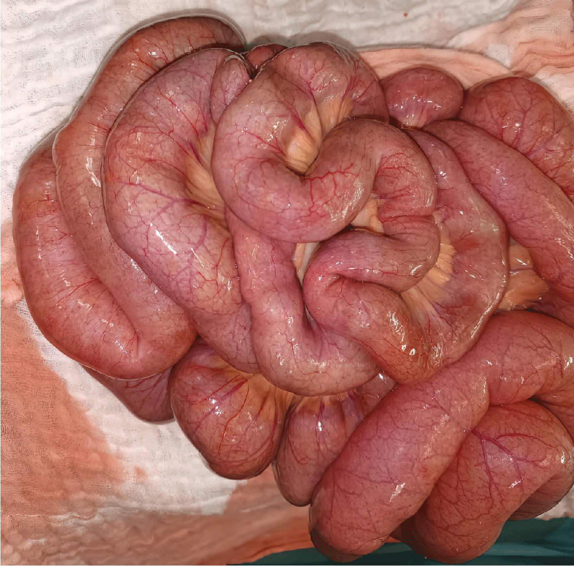
In view of the clinical symptomatology and the CT scan results, the patient underwent an exploratory laparotomy with the following findings: Absence of peritoneal effusion with the presence of a 3 cm distension of the bowel without any sign of flange, stenosis, or detectable obstacle, with the performance of multiple biopsies at the level of the greater omentum, the mesentery, and the anterior parietal peritoneum
Conclusion: Intestinal pseudo-obstruction is a rarely reported manifestation during systemic lupus erythematosus. It is a rare but potentially serious manifestation that can reveal the disease or occur during the course of the disease. The use of immunosuppressive drugs is reserved for recurrent forms or in case of failure of corticosteroids. However, recurrences are frequent. Azathioprine is an alternative therapy to control the disease.
However, recurrences are frequent. Azathioprine is an alternative therapy to control the disease.Introduction
Systemic Lupus Erythematosus (SLE) is a non-organ-specific autoimmune disease that affects women of childbearing age in preference to men and progresses in relapses.
It is a rare but potentially serious manifestation that can reveal the disease or occur during its evolution.
It is characterized by very polymorphic clinical manifestations. Lupus enteritis is one of the complications of lupus and a cause of abdominal pain syndrome [1]. It is thought to be related to digestive vasculitis. Therefore, it requires a rapid diagnostic and therapeutic approach, since the mortality associated with perforation or digestive hemorrhage is not negligible [2]. Recurrences under corticosteroids are frequent [3]. In these cases, the association of an immunosuppressant is necessary to stabilize the disease.
Patient and observation
Mrs. SQ, S, 24 years old, was diagnosed one week ago with systemic lupus erythematosus at the beginning of treatment, with a history of pancreatitis stage E, history of current illness goes back to 05 days before her admission by the installation of a sub-occlusive syndrome made of cessation of matter and gas associated with food vomiting, without externalized digestive hemorrhage, all of this evolving in a context of asphyxia and decline of general condition with cessation of matter and gas in the last 48 hours.
Evolving in a context of apyrexia and decline of the general state with cessation of matter and gas in the last 48 hours. At the clinical examination, we note on the general plan: Patient is conscious stable hemodynamic and respiratory, and with normal colored conjunctiva on the abdominal plan: Absence of a laparotomy scar with distended Abdomen, tympanic with rectal touch: no palpable mass, fingertips return soiled with normal colored stool and The rest of the somatic examination is without particularities, At the biology, we found a sedimentation rate at 80 mm at the first hour, a negative C-reactive protein (CRP), hemoglobin 7.6 g/dl with a hyperleukocytosis at: 14.520/mm3; normal renal workup with Urea: 0.41 g/L Creat: 6.1 mg/L.
Para-clinical The Abdomen without preparation showed hydroaeric levels (Figure 1) with an abdominal CT scan (Figure 2) which showed: CT scan appearance in favor of bowel obstruction with evidence of a transitional level above the umbilical: inflammatory stenosis? The spontaneously hyper-dense appearance of the colonic lumen and some ileal intestines is most probably related to a digestive hemorrhage that could be part of lupus enteritis given the patient’s past history with the spontaneously hyper-dense appearance of the colonic lumen and ileal intestines most probably related to a digestive hemorrhage. And distension of the jejunal and some ileal coves, measuring: 36mm in maximum diameter, the seat of hydroaerobic level with a transitional level above umbilical. Significant gastric distension. No parietal pneumatosis with no parietal enhancement defect.
In view of the clinical symptomatology and the CT scan results, the patient underwent an exploratory laparotomy (Figure 3) with the following findings: Absence of peritoneal effusion with the presence of a 3 cm distension of the bowel without any sign of flange, stenosis or detectable obstacle, with the performance of multiple biopsies at the level of the greater omentum, the mesentery, and the anterior parietal peritoneum.
Discussion
Gastrointestinal manifestations observed in SLE are common, in the order of 50%. They may involve any segment of the digestive tract. Oral mucosal lesions, esophageal dysmotility, exudative enteropathy, and pancreatitis are the most frequent manifestations [4]. These manifestations may be directly related to the lupus disease or secondary to the prescribed treatments.
Intestinal Pseudo-Obstruction (IOP) is rarely described in SLE with only 25 cases recently reported. IOP may be a manifestation of the disease, may occur during the course of active disease, or may even be the only manifestation of an SLE flare. Lupus enteritis is a rare complication of SLE. Its prevalence is estimated at between 0.2% and 2%. However, it is a frequent cause of abdominal pain in some series, reported in 45% - 79% of cases [5,6].
Pathophysiologically, lupus enteritis is the consequence of vasculitis of the small vessels (arterioles and venules) associating atrophy, degeneration of the media, fibrinoid necrosis, old thrombosis, phlebitis, and infiltration of the lamina propria by monocytes [7,8].
The clinical picture is that of an acute intestinal obstruction manifested by abdominal pain, nausea or vomiting, transit disorders, abdominal distension, and weight loss [9-11]. No obstruction is found, but it is the intestinal motility that is ineffective. It often associates acute abdominal pain, vomiting, diarrhea, sometimes even an occlusive syndrome, and signs of peritoneal irritation [2,7]. Our patient presented with abdominal pain, diarrhea, vomiting, and even a sub-occlusive syndrome which became occlusive after one week of evolution.
Several etiopathogenic hypotheses have been put forward to explain the intestinal motor disorder [11,12]:
- A primary neurogenic involvement;
- The existence of autoantibodies directed against the myocytes
- Intestinal vasculitis mediated by circulating immune complexes
- Congenital smooth muscle dysfunction in idiopathic forms.
The diagnosis of POI is based on clinical and imaging findings of small bowel or colonic lumen dilatation with wall thinning and multiple hydro-aerobic levels occasionally associated with bilateral ureteral dilatation and reduced bladder capacity.
Abdominal CT scan shows bowel wall thickening, cocoon images, mesenteric edema, and sometimes ascites. However, these lesions are not specific, as they can be seen in other digestive diseases [5]. The CT scan can therefore eliminate other causes of abdominal pain such as pancreatitis. Thus, the diagnosis of lupus enteritis remains a diagnosis of exclusion and requires in all cases a complete workup to eliminate other causes of abdominal pain and infectious causes [3,13]. Digestive endoscopies, when performed, usually reveal a highly edematous digestive wall and even ulcerations [3].
The manometric study shows global intestinal and gastric hypomotricity and sometimes esophageal aperistalsis, but it is more useful in other connectivities, mainly scleroderma [12,14]. There are no specific immunological markers, but anti-Ro antibody positivity has been described in 75% of cases [15]. The association of ureterohydronephrosis and cystitis [16] is found in 63% of the cases of POI related to SLE reported in the literature [17].
This dual digestive and urological involvement can be attributed to diffuse smooth muscle involvement [12]. Complications of enteritis are rare. They are related to massive hemorrhage or digestive perforation that can worsen the vital prognosis [7,13]. Radiological examinations are of great help in the diagnosis. The involvement is most often jejunal or ileal [5,6,13]. Our patient did not have any jejunal involvement, but she had anemia of 7 with CT signs of intra-luminal hemorrhage.
The evolution is often favorable after early treatment, but recurrence of the symptoms has been described as well as deaths secondary to ischemia or intestinal perforation (18%) [2].
The initial treatment of lupus enteritis is based on high doses of corticosteroids resulting in a good response. However, relapses are not uncommon, even in patients with a good initial response to this treatment [2,7]. Kim, et al. [18] showed that there were no significant differences in epidemiological, clinical, or biological data, including autoantibody profile and SLEDAI, between lupus enteritis with and without relapse. However, he noted that in patients without recurrence, the cumulative dose of prednisolone and the duration of treatment were significantly higher than in patients with recurrence.
Treatment of IOP is based on symptomatic measures combining parenteral nutrition, discontinuation of drugs that may induce or aggravate IOP, correction of fluid and electrolyte disorders (hypocalcemia or hypokalemia), antibiotic therapy aimed at the digestive flora, and intestinal motor stimulators such as erythromycin or octreotide. High-dose corticosteroid therapy seems to be the treatment of choice, but the association with immunosuppressants (azathioprine or cyclophosphamide) may be necessary for severe forms or in the presence of extra digestive manifestations, particularly renal [9]. The use of immunosuppressants in our patient was dictated by severe hematological and glomerular involvement.
The use of other therapies is sometimes necessary in case of failure of corticosteroids or in case of corticosteroid dependence. Grimbacher [8] reported a complete remission of recurrent lupus enteritis with cyclophosphamide infusions. Other therapeutics have proven to be effective such as mycophenolate mofetil, azathioprine, tacrolimus, and Rituximab [2,19,20]. Our case is characterized by frequent recurrences at high doses of corticosteroids requiring the addition of azathioprine which allowed stabilization of the disease.
Conclusion
Lupus enteritis is a rare manifestation, but one of the main causes of acute abdominal pain in SLE. Its diagnosis is based on clinical, biological, and radiological elements, after the elimination of other causes of abdominal pain. Treatment is based mainly on corticosteroids.
Intestinal Pseudo-Obstruction (IOP) is a rarely reported manifestation of Systemic Lupus Erythematosus (SLE). It is a rare but potentially serious manifestation, which may reveal the disease or occur during its course.
However, recurrences are frequent. Azathioprine is an alternative treatment that can control the disease.
Consent written
Written informed consent was obtained from the patient for the publication of this case report and accompanying images. A copy of the written consent is available for review by the Editor-in-Chief of this journal on request.
Authors’ contributions
This work was carried out in collaboration among all authors. All authors contributed to the conduct of this work. They also declare that they have read and approved the final version of the manuscript.
- Richer O, Ulinski T, Lemelle I, Ranchin B, Loirat C, Piette JC, Pillet P, Quartier P, Salomon R, Bader-Meunier B; French Pediatric-Onset SLE Study Group. Abdominal manifestations in childhood-onset systemic lupus erythematosus. Ann Rheum Dis. 2007 Feb;66(2):174-8. doi: 10.1136/ard.2005.050070. Epub 2006 Jul 3. PMID: 16818463; PMCID: PMC1798515.
- Shirai T, Hirabayashi Y, Watanabe R, Tajima Y, Fujii H, Takasawa N, Ishii T, Harigae H. The use of tacrolimus for recurrent lupus enteritis: a case report. J Med Case Rep. 2010 May 24;4:150. doi: 10.1186/1752-1947-4-150. PMID: 20497521; PMCID: PMC2887895.
- Thomas G, Ebbo M, Genot S, Bernit E, Mazodier K, Veit V, Lagrange X, Heyries L, Kaplanski G, Schleinitz N, Harlé JR. L'entérite : une manifestation peu fréquente et corticosensible du lupus érythémateux systémique [Lupus enteritis: an uncommon manifestation of systemic lupus erythematosus with favourable outcome on corticosteroids]. Rev Med Interne. 2010 Jul;31(7):493-7. French. doi: 10.1016/j.revmed.2010.01.006. Epub 2010 May 14. PMID: 20471141.
- Perlemuter G, Cacoub P, Wechsler B, Hausfater P, Piette JC, Couturier D, Chaussade S. Pseudo-obstruction intestinale chronique secondaire aux connectivites [Chronic intestinal pseudo-obstruction secondary to connective tissue diseases]. Gastroenterol Clin Biol. 2001 Mar;25(3):251-8. French. PMID: 11395671.
- Lee CK, Ahn MS, Lee EY, Shin JH, Cho YS, Ha HK, Yoo B, Moon HB. Acute abdominal pain in systemic lupus erythematosus: focus on lupus enteritis (gastrointestinal vasculitis). Ann Rheum Dis. 2002 Jun;61(6):547-50. doi: 10.1136/ard.61.6.547. PMID: 12006332; PMCID: PMC1754133.
- Byun JY, Ha HK, Yu SY, Min JK, Park SH, Kim HY, Chun KA, Choi KH, Ko BH, Shinn KS. CT features of systemic lupus erythematosus in patients with acute abdominal pain: emphasis on ischemic bowel disease. Radiology. 1999 Apr;211(1):203-9. doi: 10.1148/radiology.211.1.r99mr17203. PMID: 10189472.
- Sultan SM, Ioannou Y, Isenberg DA. A review of gastrointestinal manifestations of systemic lupus erythematosus. Rheumatology (Oxford). 1999 Oct;38(10):917-32. doi: 10.1093/rheumatology/38.10.917. PMID: 10534541.
- Grimbacher B, Huber M, von Kempis J, Kalden P, Uhl M, Köhler G, Blum HE, Peter HH. Successful treatment of gastrointestinal vasculitis due to systemic lupus erythematosus with intravenous pulse cyclophosphamide: a clinical case report and review of the literature. Br J Rheumatol. 1998 Sep;37(9):1023-8. doi: 10.1093/rheumatology/37.9.1023. PMID: 9783772.
- Ceccato F, Salas A, Góngora V, Ruta S, Roverano S, Marcos JC, Garcìa M, Paira S. Chronic intestinal pseudo-obstruction in patients with systemic lupus erythematosus: report of four cases. Clin Rheumatol. 2008 Mar;27(3):399-402. doi: 10.1007/s10067-007-0760-5. Epub 2007 Oct 16. PMID: 17938989.
- Alexopoulou A, Andrianakos A, Dourakis SP. Intestinal pseudo-obstruction and ureterohydronephrosis as the presenting manifestations of relapse in a lupus patient. Lupus. 2004;13(12):954-6. doi: 10.1191/0961203304u1064cr. PMID: 15645752.
- Hill PA, Dwyer KM, Power DA. Chronic intestinal pseudo-obstruction in systemic lupus erythematosus due to intestinal smooth muscle myopathy. Lupus. 2000;9(6):458-63. doi: 10.1191/096120300678828505. PMID: 10981652.
- Perlemuter G, Cacoub P, Wechsler B, Hausfater P, Piette JC, Couturier D, Chaussade S. Pseudo-obstruction intestinale chronique secondaire aux connectivites [Chronic intestinal pseudo-obstruction secondary to connective tissue diseases]. Gastroenterol Clin Biol. 2001 Mar;25(3):251-8. French. PMID: 11395671.
- Taourel PG, Deneuville M, Pradel JA, Régent D, Bruel JM. Acute mesenteric ischemia: diagnosis with contrast-enhanced CT. Radiology. 1996 Jun;199(3):632-6. doi: 10.1148/radiology.199.3.8637978. PMID: 8637978.
- Perlemuter G, Chaussade S, Wechsler B, Cacoub P, Dapoigny M, Kahan A, Godeau P, Couturier D. Chronic intestinal pseudo-obstruction in systemic lupus erythematosus. Gut. 1998 Jul;43(1):117-22. doi: 10.1136/gut.43.1.117. PMID: 9771415; PMCID: PMC1727185.
- Leclair MA, Plaisance M, Balfour S, Touchette M. Chronic intestinal pseudoobstruction in systemic lupus erythematosus. Can J Gen Inter Med 2008; 3: 18–20.
- Pardos-Gea J, Ordi-Ros J, Selva A, Perez-Lopez J, Balada E, Vilardell M. Chronic intestinal pseudo-obstruction associated with biliary tract dilatation in a patient with systemic lupus erythematosus. Lupus. 2005;14(4):328-30. doi: 10.1191/0961203304lu2047cr. PMID: 15864921.
- Chen YY, Yen HH, Hsu YT. Intestinal pseudo-obstruction as the initial presentation of systemic lupus erythematosus: the need for enteroscopic evaluation. Gastrointest Endosc. 2005 Dec;62(6):984-7. doi: 10.1016/j.gie.2005.08.010. PMID: 16301053.
- Kim YG, Ha HK, Nah SS, Lee CK, Moon HB, Yoo B. Acute abdominal pain in systemic lupus erythematosus: factors contributing to recurrence of lupus enteritis. Ann Rheum Dis. 2006 Nov;65(11):1537-8. doi: 10.1136/ard.2006.053264. PMID: 17038460; PMCID: PMC1798347.
- Kishimoto M, Nasir A, Mor A, Belmont HM. Acute gastrointestinal distress syndrome in patients with systemic lupus erythematosus. Lupus. 2007;16(2):137-41. doi: 10.1177/0961203306075739. PMID: 17402371.
- Janssens P, Arnaud L, Galicier L, Mathian A, Hie M, Sene D, Haroche J, Veyssier-Belot C, Huynh-Charlier I, Grenier PA, Piette JC, Amoura Z. Lupus enteritis: from clinical findings to therapeutic management. Orphanet J Rare Dis. 2013 May 3;8:67. doi: 10.1186/1750-1172-8-67. PMID: 23642042; PMCID: PMC3651279.

Article Alerts
Subscribe to our articles alerts and stay tuned.
 This work is licensed under a Creative Commons Attribution 4.0 International License.


 Save to Mendeley
