Archives of Community Medicine and Public Health
Polar and extrahilar renal arteries: A case report
Yoanna Vladimirova Tivcheva1,2*, Mihail Angelov1, Nikolai Krastev1, Cvetomir Kirilov1, Dimo Krastev1-3, L Jelev1-3 and A Apostolov3
2Medical College “Jordanka Filaretova”, Medical University of Sofia, 3 Jordanka Filaretova Str., 1606 Sofia, Bulgaria
3Faculty of Public Health, Health Care and Sport, South-West University “Neofit Rilski”, 66 Ivan Mihailov Str., 2700 Blagoevgrad, Bulgaria
Cite this as
Tivcheva YV, Angelov M, Krastev N, Kirilov C, Krastev D, et al. (2023) Polar and extrahilar renal arteries: A case report. Arch Community Med Public Health 9(1): 001-003. DOI: 10.17352/2455-5479.000193Copyright License
© 2023 Tivcheva YV, et al. This is an open-access article distributed under the terms of the Creative Commons Attribution License, which permits unrestricted use, distribution, and reproduction in any medium, provided the original author and source are credited.Introduction: The vascular system has a high frequency of variations, which are of interest to both anatomists and clinicians, as well as surgeons. The renal vasculature is quite variable and given the significant number of variations, the latter has proven difficult to classify. The conflicting terminology is often the cause of a poor understanding of the clinical implications of the presence of such variations. We present a case of bilateral accessory arteries, which can be classified as polar and extrahilar.
Background: Variants of the renal artery are a common finding with additional vessels in up to 30% of cases. The supernumerary arteries are of end type and often enter the kidney outside the hilum. The arteries that enter the kidney in its upper or lower pole are referred to as polar arteries.
Case report: During a routine dissection of a 73-year-old, female, formalin-fixed cadaver at the department of Anatomy, Histology and Embryology at the Medical University of Sofia, we discovered a right inferior polar artery and a left extrahilar renal artery, both originating from the abdominal aorta. The right kidney was located at the level of L1- L2.
Conclusion: Accessory renal vessels have been an object of multiple cadaveric and in vivo studies. The terminology and classification of such variations in regard to their origin, course, and site of entrance in the kidney are conflicting and often prove inadequate to convey the clinical and surgical importance of their presence. Knowledge of such variants is of great significance when performing an explorative laparotomy, kidney transplantation, and assessing kidney injury. Such vessels are as well associated with cases of hypertension, hydronephrosis and other conditions.
Introduction
The vascular system has a high frequency of variations, which are of interest to both anatomists and clinicians, as well as surgeons. The renal vasculature is quite variable and given the significant number of variations, the latter has proven difficult to classify. The conflicting terminology is often the cause of a poor understanding of the clinical implications of the presence of such variations. We present a case of bilateral accessory arteries, which can be classified as polar and extrahilar.
Background
The human kidneys, normally located in the retroperitoneal space, receive blood supply from a pair of renal arteries, the origin of which is determined by the skeletotopy of the organ. As kidneys are normally located at the level of the L1-L2 vertebral body, this is the level of the respective arteries. The renal artery enters the kidney through the hilum and further branches out into segmental arteries, which are considered anatomically terminal. An accessory artery is supplementary to the renal artery that occurs in approximately 30% of people and also takes part in supplying the kidney with blood. It can enter the kidney through the hilum or more commonly through either the superior or the inferior pole of the kidney. In those cases, it is considered a polar artery and is associated with additional clinical implications.
Case report
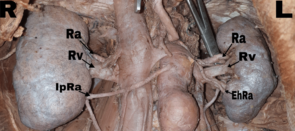
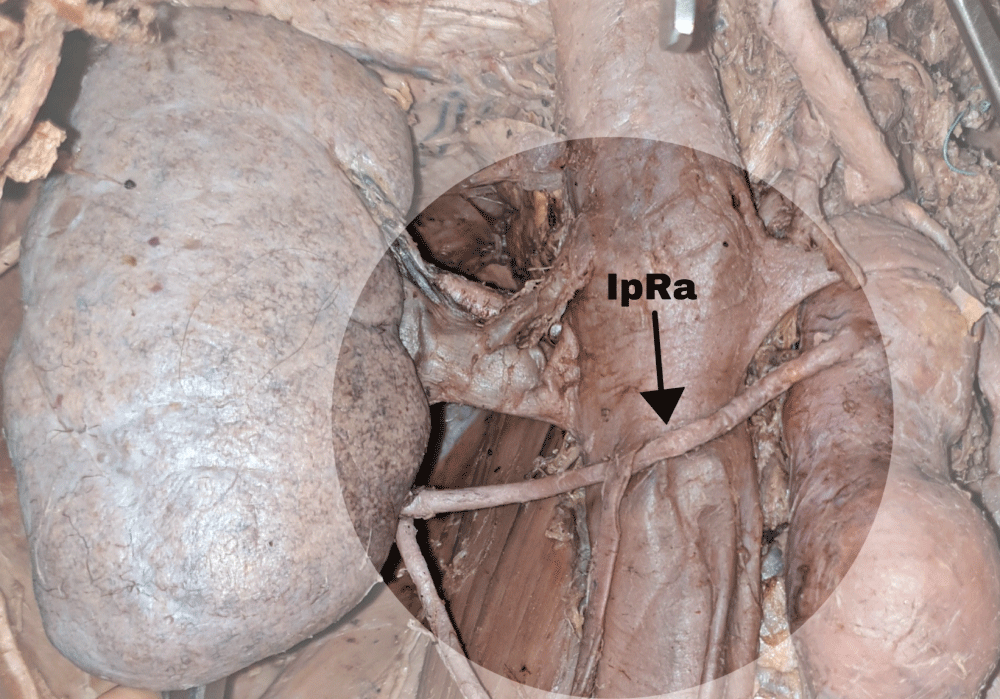
During a routine dissection of a 73-year-old, female, formalin-fixed cadaver at the department of Anatomy, Histology, and Embryology at the Medical University of Sofia, we discovered a right inferior polar artery and a left accessory renal artery, both originating from the abdominal aorta (Figure 1). The right kidney was located at the level of L1- L2 (Figure 2). Upon closer inspection of the hilum, the vessels had a normal distribution with the renal vein to the front and the renal artery behind it. An accessory renal artery was also found, emerging about 30 mm under the superior mesenteric artery and descending obliquely, and entering the kidney medially, outside of the hilum at the lower pole. The left kidney was at the level of Th12 - L1. Upon decapsulation and further dissection of the vessels, an accessory renal artery was found. The latter was emerging closely under the superior mesenteric artery, descending obliquely, crossing over the renal vein and entering the kidney medially, outside the hilum, above the lower pole. The artery was further found to supply the lower segment of the kidney (Figure 3).
Discussion
During a routine dissection of a 73-year-old, female, formalin-fixed cadaver at the department of Anatomy, Histology, and Embryology at the Medical University of Sofia, we discovered a right inferior polar artery and a left accessory renal artery, both originating from the abdominal aorta (Figure 1). The right kidney was located at the level of L1- L2 (Figure 2). Upon closer inspection of the hilum, the vessels had a normal distribution with the renal vein to the front and the renal artery behind it.
The anatomy of the kidney and its respective blood supply have been an object of multiple studies both cadaveric and in vivo. The renal artery varies in origin, course, and also in a number of arteries.
The first records of multiple renal arteries date back to 1552 by Eustachius. The presence of multiple renal arteries is by far the most common variation, with up to six separate arteries. In those cases, the renal artery of the biggest caliber and typical anatomical position is referred to as the main renal artery. Up to 30% of individuals have additional renal vessels. In 16% of the cases, the arteries vary between the kidneys of the same individual [1]. The accessory vessels emerge predominantly from the aorta, but also the superior mesenteric artery, the renal artery. There are rare cases of accessory vessels branching from the suprarenal, inferior mesenteric, common, and internal iliac arteries [2].
Further, the vast number of variations has been classified multiple times, often with conflicting terminology. Eisendrath offers a classification of five types in 1910 [3], based on origin, site of entrance in the kidney, as well as the number of vessels. Even as early as that the terminology proves inaccurate as such vessels are anatomically terminal or end arteries and should not be referred to as “additional”. Graves uses the terms accessory and aberrant as interchangeable [4], yet many authors consider aberrant vessels as vessels entering the kidney outside the hilum [5]. In many cases additional vessels are referred to as supernumerary or supplemental [6,7]. We consider such a term appropriate for the group of variations, but we will refer to arteries entering the pole of the kidney as polar and to arteries entering outside the hilum, but not at the pole as extrahilar, as proposed by Sampaio and Passos [8].
The development of such variation is associated with the complex vasculature of the embryological kidney. The mesonephric vessels, one of which later gives rise to the renal artery, can persist as more than one, which eventually results in multiple renal arteries [9]. The presence of such vessels takes part in the pathogenesis of multiple conditions. Clinical findings of increased renin-angiotensin-aldosterone system activity have been associated with multiple or supernumerary renal arteries. Therefore individuals carrying those variants are at risk for secondary or renal hypertension [10,11]. It is worth mentioning that such patients are candidates for renal denervation, which can prove difficult in the presence of variant vasculature [12]. Supernummeral arteries crossing the ureteropelvic junction and specifically inferior pole arteries have been associated with up to 7% of the cases of ureteropelvic obstruction and further hydronephrosis [13,14]. The radiological findings of course are also quite specific in patients with such variations. Alongside other procedures in the retroperitoneal space that can be complicated by variant vasculature of the kidney, kidney transplantation is by far the most documented. Not only is the procedure itself challenging but also the graft viability and graft function differ when compared to single artery transplant. Though the long-term results are similar and multiple artery grafts are not contraindicated in transplant, the data has shown the importance and specifics of the procedure in the presence of multiple arteries [15].
Conclusion
The variations of the renal arteries are a common finding. A good understanding of the vascular anatomy of the kidney and its variations is fundamental to good surgical and clinical practice. It is of great significance when performing an explorative laparotomy, kidney transplantation, interpreting angiographies and other radiological data involving the kidneys, and assessing kidney injury. A comprehensive review of the renal arteries variations clearly shows the need for a more structured classification, as the clinical and surgical implications of the presence of such variants can significantly change the outcome for patients carrying them.
- Priti LM. Renal Arteries; 2016. B Bergman's Comprehensive Encyclopedia of Human Anatomic Variation 682-693. https://doi.org/10.1002/9781118430309.ch55
- MERKLIN RJ, MICHELS NA. The variant renal and suprarenal blood supply with data on the inferior phrenic, ureteral and gonadal arteries: a statistical analysis based on 185 dissections and review of the literature. J Int Coll Surg. 1958 Jan;29(1 Pt 1):41-76. PMID: 13502578.
- Eisendrath DN, Strauss DC. The surgical importance of accessory renal arteries. JAMA.1910; 55(16):1375-1377. doi:10.1001/jama.1910.04330160043015
- Graves FT. The aberrant renal artery. J Anat. 1956 Oct;90(4):553-8. PMID: 13366870; PMCID: PMC1244871.
- Standring S. ed. Gray’s Anatomy. The Anatomical Basis of Clinical Practice. 40th Ed., Edinburg, Churchill & Livingstone. 2008; 1231-1233.
- Anson BJ, Richardson GA, Minear WL. Variations in the number and arrangements of the renal blood vessels, a study of the blood supply of four hundred kidneys. Journal of Urology. 1936; 36: 211-219.
- Talovic E, Kulenovic A, Voljevica A, Kapur E. Review of supernumerary renal arteries by dissection method. Acta Medica Academica. 2007; 36:59-69.
- Sampaio FJ, Passos MA. Renal arteries: anatomic study for surgical and radiological practice. Surg Radiol Anat. 1992;14(2):113-7. doi: 10.1007/BF01794885. PMID: 1641734.
- Felix W. Mesonephric arteries (aa. mesonephric) In Keibel F, Mall FP, editors. Manual of Human Embryology. 22. Philadelphia, Pa, USA: Lippincott; 1912.
- Glodny B, Cromme S, Reimer P, Lennarz M, Winde G, Vetter H. Hypertension associated with multiple renal arteries may be renin-dependent. J Hypertens. 2000 Oct;18(10):1437-44. doi: 10.1097/00004872-200018100-00011. PMID: 11057431.
- Chan PL, Tan FHS. Renin dependent hypertension caused by accessory renal arteries. Clin Hypertens. 2018 Nov 1;24:15. doi: 10.1186/s40885-018-0100-x. PMID: 30410790; PMCID: PMC6211501.
- Verloop WL, Vink EE, Spiering W, Blankestijn PJ, Doevendans PA, Bots ML, Vonken EJ, Voskuil M. Renal denervation in multiple renal arteries. Eur J Clin Invest. 2014 Aug;44(8):728-35. doi: 10.1111/eci.12289. PMID: 24931208.
- Johnston JH. The pathogenesis of hydronephrosis in children. Br J Urol. 1969 Dec;41(6):724-34. doi: 10.1111/j.1464-410x.1969.tb09985.x. PMID: 5359497.
- NIXON HH. Hydronephrosis in children; a clinical study of seventy-eight cases with special reference to the role of aberrant renal vessels and the results of conservative operations. Br J Surg. 1953 May;40(164):601-9. doi: 10.1002/bjs.18004016416. PMID: 13059346.
- Zorgdrager M, Krikke C, Hofker SH, Leuvenink HG, Pol RA. Multiple Renal Arteries in Kidney Transplantation: A Systematic Review and Meta-Analysis. Ann Transplant. 2016 Jul 29;21:469-78. doi: 10.12659/aot.898748. PMID: 27470979.
Article Alerts
Subscribe to our articles alerts and stay tuned.
 This work is licensed under a Creative Commons Attribution 4.0 International License.


 Save to Mendeley
